Pregnancy scanning
Complete Ultrasound Services
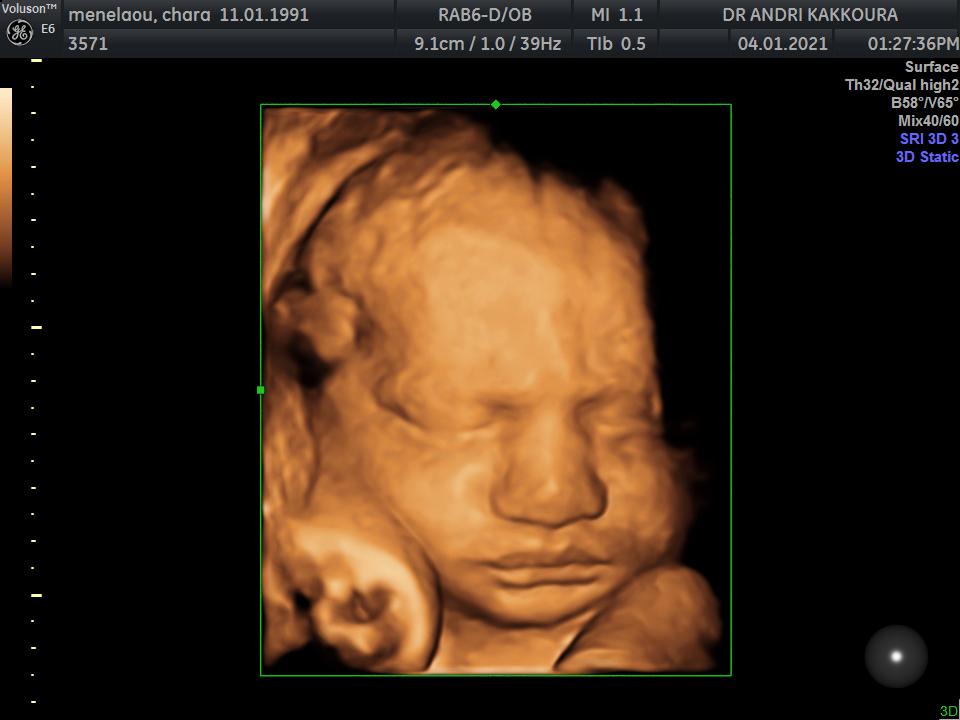
Dr. Kakkoura offers both routine ultrasound checkups as well as advanced diagnostic pregnancy ultrasound evaluations. She is renowned for her expertise in the field of ultrasound imaging in both early as well as advanced pregnancy. She adopts the latest available technology, and her ultrasound equipment is state of the art and includes 4D imaging.
Thanks to Dr. Kakkoura’s specialization in pregnancy scanning, it is possible during the same appointment to conduct both your own obstetric examination as well as the detailed fetal ultrasound are seen, scanned, and diagnosed all during the same appointment.
Because of our Doctor specialization in pregnancy scanning, you can have during your entire pregnancy a one-stop ultrasound scanning service, whereby you are seen, scanned and diagnosed all during the same appointment.
What you need to know when conducting ultrasound assessments
Ultrasound scanning is performed using ultrasound waves, which cannot be heard or felt. The technique provides an image of the fetus and placenta. Ultrasounds arep harmless to both the mother and the embryo and as such can be repeated as frequently as necessary. Ultrasound scanning represents a very valuable diagnostice tool during pregnancy
Routine Pregnancy Scanning
The scans conducted during the early stages of pregnancy are primarily used to:
- Establish single, twin, or multiple pregnancy
- Diagnose the viability of the fetus by detection of cardiac function
- Confirm the stage of the pregnancy through measuring the fetus length
- Detect or rule out an ectopic pregnancy
The first scan, done in the 6th week of pregnancy, is a transvaginal ultrasound (through the vagina) where the fetal sac is visible. Cardiac function is detected after the 6+ week.
As of the 8th-9th week of pregnancy, it is possible to conduct transabdominal ultrasound scans (over the abdomen).
In the early stages of pregnancy, the ultrasound scans also help determine the right time to conduct the nuchal translucency scan.
The nuchal translucency scan is conducted between 11 weeks and 13 weeks plus 6 days o pregnancy. When evaluated in combination with the blood tests conducted during the 9th – 10th week, it is possible to assess the risk of developing chromosomal abnormalities.
All fetuses collect fluid at the back of their neck. The more fluid collected, the higher the risk of any chromosomal abnormalities. The ultrasound is used to measure the thickness of this fluid.
During this scan, the doctor also checks the basic anatomy of the embryo and the presence or not of the nasal bone. Furthermore, a specific doppler ultrasound is conducted (ductus venosus) to evaluate the embryo’s blood flow to predict possible heart conditions. Finally, the Doppler measurement of the mother’s uterine arteries determines the possible risk to develop preeclampsia in later stages of the pregnancy.
Between 20 weeks and 24 weeks of pregnancy, the anomaly scan is performed to detect the presence of any fetal anatomic abnormalities. During this scan, all fetal organs (abdomen, bones, brain, face, heart, kidneys and spinal cord) are thoroughly inspected as well as the health and position of the placenta. Furthermore, the volume of amniotic fluid surrounding the embryo is checked.
The length of the cervix is also measured to determine if there is a risk for premature birth. Finally, through a doppler measurement ultrasound of the uterine arteries, it is possible to determine the risk of preeclampsia at a later stage of the pregnancy or residual fetal development.
During the 3rd trimester, between the 28th and 34th week, the ultrasound scan determines the baby’s measurements and overall wellbeing and health of the embryo.
Call Us
+357 22 476889
Reach Us
American Medical Center
215, Spyrou Kyprianou Avenue, 2047, Strovolos, Nicosia, Cyprus
Open Hours
Mon, Wed, Fri: 8:00 - 20:00
Tue: 8:00 - 17:00
Thu: 8:00 - 14:00